Scaphoid x-rays are indicated for a variety of settings including:
- wrist trauma
- bony tenderness at the anatomical snuffbox
- suspected fracture
- obvious deformity
- non-traumatic wrist pain
Projections
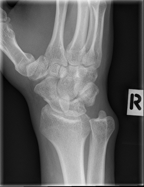
Standard projections
- PA view
- ulnar deviation to remove the scaphoid from the radius and present its axis longitudinally
- the best view to inspect the joint spaces of the carpal bones and the distal radio-ulnar joint

- PA view angled
- ulnar deviation to remove the scaphoid from the radius and present its axis longitudinally
- tube angulation to present the scaphoid en face

- oblique view
- external oblique projection
- lateral view
- projection 90° to the PA view
- demonstrates multiple carpal bones overlapping
- the essential view to assessing the alignment of the radius, lunate, and capitate in the setting of a suspected dislocation

Modified trauma projections
- horizontal beam lateral view
- modified lateral projection that requires little to no patient movement
- produces a diagnostic lateral projection without risking patient pain

Additional projections
- clenched fist view
- used for suspected scapholunate dissociation

Mayo classification:
Three types according to anatomic location of the fracture line:
- middle (70%)
- distal (20%)
- proximal (10%)
Fractures of the distal third are further divided into distal articular surface or the distal tubercle fractures:
- distal tubercle third fracture
- distal articular surface fracture
- distal third fracture
- middle third fracture
- proximal third fracture
Management options can broadly be divided into:
- immobilisation with cast application
- internal fixation for displaced fragments, usually with a headless self-compressing screw
- non-union can be managed with internal fixation and bone grafting
Factors affecting prognosis:
- location 9
- distal pole: excellent likelihood of union (~100%)
- waist: ~10-20% chance of non-union
- proximal pole: ~30-40% chance of non-union
- vertically oriented fracture line
- fragment displacement of greater than 1mm
- ligamentous instability: increased scapholunate angle (i.e. >60º or radiolunate or capitolunate angle >15º)
The major complication of scaphoid fractures is non-union or malunion leading to instability and secondary osteoarthritic change. Hence surgical treatment for displaced fractures or angulation.
A number of other specific complications are encountered from time to time:
- avascular necrosis: Preiser disease
- this usually involves the proximal portion as a result of blood supply to the scaphoid entering relatively distally
- SNAC wrist: scaphoid non union advanced collapse
- SLAC wrist: scapholunate advanced collapse
*Source extracted from :https://radiopaedia.org/articles/scaphoid-fracture